| 一般情况 | |
|---|---|
 | 品种:孟加拉猫 |
| 年龄:2岁 | |
| 性别:雄 | |
| 是否绝育:是 | |
| 诊断:食道狭窄 | |
01 主诉及病史
固体食物吞咽困难和慢性反胃超过5个月。
在当地一家医院接受了约2个月的胃炎治疗,但临床症状未见好转。无其他临床病史或治疗史。
02 检查
身体状况评分为3分(满分9分),收缩压120 mmHg,呼吸频率40次/分,直肠体温38.6℃,心率160次/分,听诊无心律失常。血液检查未发现明显异常。
丙氨酸氨基转移酶活性43 U/L(20-107)
天冬氨酸氨基转移酶活性25 U/L(6-44)
碱性磷酸酶活性80 U/L(23-107)。
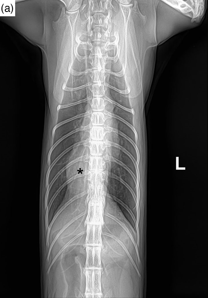
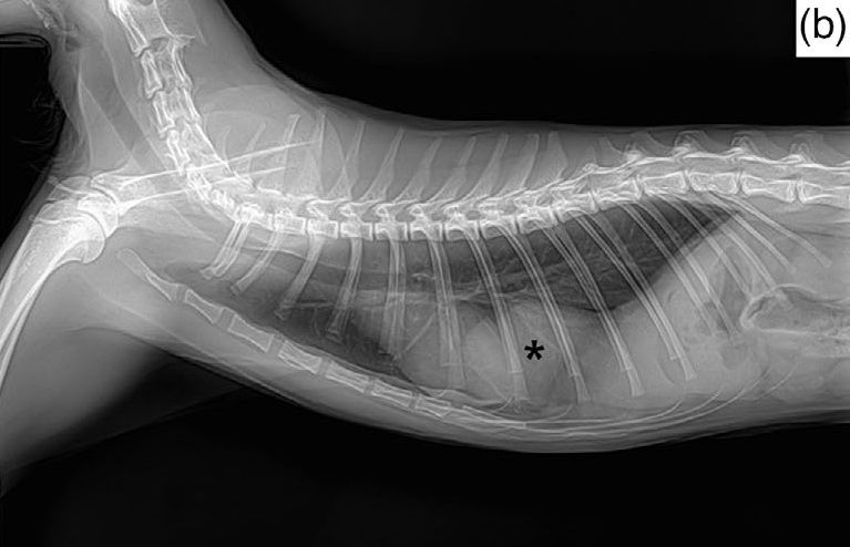
胸片显示胸腔内从腹部突起一个均质软组织不透明肿块(3.5×4.0 cm),遮挡了尾腔静脉,并与心脏右心尖相邻。胃肠移位不明显,但由于膈角移位,怀疑食道和贲门之间的夹角增大(下图)。
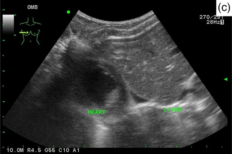
超声检查在经肝切面上发现了一个突出的肝叶,周围有一层从横膈向胸腔方向延伸的高回声内膜(下图)。根据这些检查结果,诊断为胸腹膈疝。
03 治疗
使用直径9.0 mm内窥镜进行食道镜检查,以确认慢性反流的原因。检查发现食道远端有食道狭窄,估计直径约2 mm,经内镜球囊扩张后,狭窄得以缓解(下图)。

将球囊扩张导管(5 Fr,4 mm)置于狭窄处的中心位置,手动充气60秒。移除球囊扩张导管后,仔细检查了有轻微出血的狭窄区域,未发现先天性穿孔。在扩张后的狭窄部位注射了曲安奈德,以防止狭窄形成。
术后连续14天使用雷尼替丁(3.5 mg/kg,q12h)、蔗糖酸钠(0.5 g,q12h)和甲氧氯普胺(0.5 mg/kg,q12h)来处理扩张后的狭窄和食道炎。喂食方案是罐头与水混合喂食。慢性反胃和吞咽困难等临床症状立即得到缓解。
04 预后
食道球囊扩张术后病情稳定,但两个月后吞咽困难和反胃症状再次出现。对该猫进行了CT检查,观察到了真性胸腹膈疝,右侧肝叶从膈肌中央肌腱处疝入胸腔(下图)。
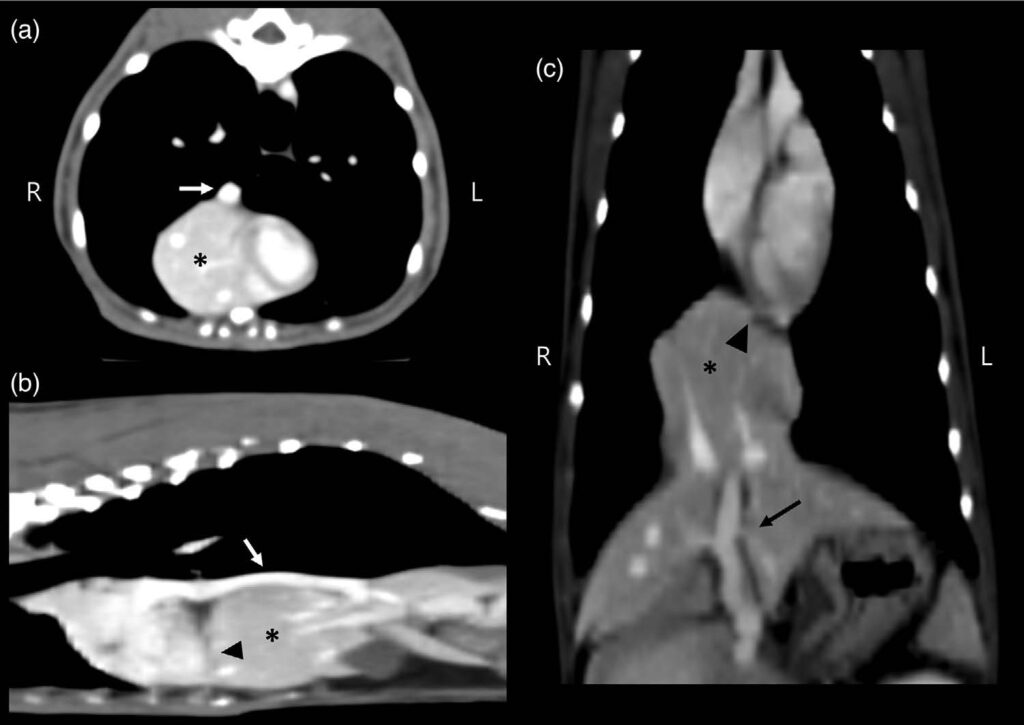
食管下括约肌有一个壁层增厚的食管狭窄(下图)。

由于怀疑食管狭窄复发是导致临床症状的原因,因此进行了第二次食管内窥镜检查和球囊扩张术。但由于主人拒绝,没有对真性膈疝进行手术修补。术后症状有所缓解,病情也保持稳定,直到约6个月后失去随访。
05 讨论
食道狭窄的定义是由纤维化或疤痕导致的食道管腔狭窄,可由机械损伤、先天性狭窄和化学损伤引起。它一般是由胃食管反流引起的食管炎所致[1,2]。临床症状包括反流、唾液分泌过多和体重减轻,严重程度取决于狭窄的位置或直径[1]。
真性胸腹膈疝是一种先天性膈肌缺损,是指腹膜包裹的腹腔脏器向胸腔疝出[3-5]。真性胸腹膈疝通常是在胸部放射线检查中偶然发现的,因为它们很少出现临床症状[3-5]。
在人类中,先天性膈疝可能伴有胃肠道合并症,尤其是胃食管反流[6-8]。胃食管反流是先天性膈疝的常见并发症,其原因包括食管运动障碍、膈嵴异常和腹腔内压力过高[8-11]。
虽然先天性膈疝的病因和病理学过程是多因素的,但胃食管交界处的移位可导致胃食管反流[12-14]。因此,膈肌缺损的猫可能会因为胃食管反流导致反复性食管炎而发生食管狭窄。本病例报告了一例猫食道狭窄病例,其原因可能是膈疝引起的胃食管反流。
胃食管反流是进餐后食管下括约肌松弛导致的一种现象。食管炎可继发于胃食管反流,但由于对猫的胃食管反流了解甚少,因此猫很少被诊断出来[15-18]。在本病例中,食管壁增厚和狭窄的胃食管异常表现与膈疝同时出现。
食管壁增厚通常由炎症、肿瘤或感染引起。由于食管壁增厚不易通过内镜或食管造影诊断,因此CT对于鉴别诊断非常重要[19]。内窥镜检查有助于区分不同的病因,如狭窄、食管炎、粘膜溃疡和肿瘤[19]。
在本病例中,CT发现食道壁增厚,内镜检查发现食道狭窄。根据这些发现,食管壁增厚和狭窄被怀疑是由炎症引起的,如食管炎,这通常是由胃食管反流引起的,而肿瘤的可能性较小,尽管由于穿孔的风险而没有进行腔内活检。
膈疝可以在没有明显临床症状的情况下发生,可能是先天性的,也可能是外伤所致。猫最常见的先天性膈疝是腹膜心包疝[20,21]。这种情况是由于发育过程中解剖结构的缺陷导致心包腔和腹腔相通[22]。
在放射学检查中,一些发现,如心脏轮廓的整体增大和心脏腰部的缺失,可能预示着腹膜心包疝,而超声可进一步确定心包内是否存在腹腔器官[22,23]。此外,CT可将腹膜心包疝与真性胸腹膈疝区分开来,这种鉴别对于制定适当的治疗方案非常重要。
食道狭窄的内窥镜球囊扩张术是一种非手术治疗方法,两到四次扩张手术可使猫的临床症状得到缓解[1]。由于内窥镜球囊扩张术后反流症状立即得到改善,本病例中临床症状的主要原因可能就是食道狭窄。
尽管情况有所改善,但食管狭窄复发的可能性依然存在,尤其是在胃食管反流、慢性呕吐和食管炎等因素与真性胸腹膈疝有关的情况下。遗憾的是,本病例未能进行手术矫正膈疝,也没有进行后续的长期随访。为了更全面地了解类似病例中膈疝与反流之间的关联,今后的研究可能会从这方面着手。
这是首例报道的猫真性胸腹膈疝和食道狭窄伴慢性反流的病例。猫食道狭窄的病因是真性胸腹膈疝引起的胃食道反流。在膈疝伴有反流的病例中,细致的诊断方法对于确定受累的解剖结构非常重要。
参考文献
[1] Glazer, A., & Walters, P. (2008). Esophagitis and esophageal strictures. Compendium (Newtown, Pa.), 30, 281–292.
[2] Tan, D. K., Weisse, C., Berent, A., & Lamb, K. E. (2018). Prospective evaluation of an indwelling esophageal balloon dilatation feeding tube for treatment of benign esophageal strictures in dogs and cats. Journal of Veterinary Internal Medicine, 32, 693-700.
[3] Cariou, M. P., Shihab, N., Kenny, P., & Baines, S. J. (2009). Surgical management of an incidentally diagnosed true pleuroperitoneal hernia in a cat. Journal of Feline Medicine and Surgery, 11, 873–877.
[4] Pilli, M., Özgencil, F. E., Seyrek-Intas, D., Gültekin, C., & Turgut, K. (2020). Pleuroperitoneal true diaphragmatic hernia of the liver in a cat. Tierarztl Prax Ausg K Kleintiere Heimtiere, 48, 292-296.
[5] Voges, A. K., Bertrand, S., Hill, R. C., Neuwirth, L., & Schaer, M. (1997). True diaphragmatic hernia in a cat. Veterinary Radiology & Ultrasound, 38, 116–119.
[6] Marseglia, L., Manti, S., D’Angelo, G., Gitto, E., Salpietro, C., Centorrino, A., Scalfari, G., Santoro, G., Impellizzeri, P., & Romeo, C. (2015). Gastroesophageal reflux and congenital gastrointestinal malformations. World Journal of Gastroenterology, 21, 8508–8515.
[7] Nagaya, M., Akatsuka, H., & Kato, J. (1994). Gastroesophageal reflux occurring after repair of congenital diaphragmatic hernia. Journal of Pediatric Surgery, 29, 1447–1451.
[8] Sigalet, D. L., Nguyen, L. T., Adolph, V., Laberge, J. M., Hong, A. R., & Guttman, F. M. (1994). Gastroesophageal reflux associated with large diaphragmatic hernias. Journal of Pediatric Surgery, 29, 1262–1265.
[9] Stolar, C. J., Levy, J. P., Dillon, P. W., Reyes, C., Belamarich, P., & Berdon, W. E. (1990). Anatomic and functional abnormalities of the esophagus in infants surviving congenital diaphragmatic hernia. American Journal of Surgery, 159, 204–207.
[10] Su, W., Berry, M., Puligandla, P. S., Aspirot, A., Flageole, H., & Laberge, J. M. (2007). Predictors of gastroesophageal reflux in neonates with congenital diaphragmatic hernia. Journal of Pediatric Surgery, 42, 1639–1643.
[11] Schmittenbecher, P. P. (1990). Postoperative follow‐up of cardia function in newborn infants after esophageal, diaphragmatic and abdominal wall abnormalities. Z Kinderchir, 45, 278-281.
[12] Ellison, G. W., Lewis, D. D., Phillips, L., & Tarvin, G. B. (1987). Esophageal hiatal hernia in small animals: Literature review and a modified surgical technique. Journal of the American Animal Hospital Association, 23, 391–399.
[13] Katsianou, H., Svoronou, M., & Papazoglou, L. G. (2014). Current views regarding hiatal hernia in dogs and cats. Hellenic Journal Companion Animal Medical, 3, 22–39.
[14] Sivacolundhu, R. K., Read, R. A., & Marchevsky, A. M. (2002). Hiatal hernia controversies—A review of pathophysiology and treatment options. Australian Veterinary Journal, 80, 48–53.
[15] Gaschen, F. (2015). Disorders of esophageal, gastric, and intestinal motility in cats. In August’s consultations in feline internal medicine (Vol. 7, pp. 117). Saunders.
[16] Han, E. (2003). Diagnosis and management of reflux esophagitis. Clinical Techniques in Small Animal Practice, 18, 231–238.
[17] Steiner, J. M. (2010). Small animal gastroenterology. Schlütersche.
[18] Samadi, F., Levine, M. S., Rubesin, S. E., Katzka, D. A., & Laufer, I. (2010). Feline esophagus and gastroesophageal reflux. AJR American Journal of Roentgenology, 194, 972–976.
[19] Barlow, J. (2011). Esophageal mural thickening. In Pearls and pitfalls in thoracic imaging: Variants and other difficult diagnoses (pp. 100–103). Cambridge University Press.
[20] MacDonald, K. (2016). Pericardial diseases. In S. J. Ettinger, (Ed.), Textbook of veterinary internal medicine (8th ed, Vol. 2, pp. 3141–3163). Saunders.
[21] Neiger, R. (1996). Peritoneopericardial diaphragmatic hernia in cats. Compendium on Continuing Education for the Practicing Veterinarian, 18, 461–468.
[22] Evans, S. M., & Biery, D. N. (1980). Congenital peritoneopericardial diaphragmatic hernia in the dog and cat: A literature review and 17 additional case histories. Journal Veterinary Radiology, 21, 108–116.
[23] Burns, C. G., Bergh, M. S., & McLoughlin, M. A. (2013). Surgical and nonsurgical treatment of peritoneopericardial diaphragmatic hernia in dogs and cats: 58 cases (1999–2008). Journal of the American Veterinary Medical Association, 242(5), 643–650.