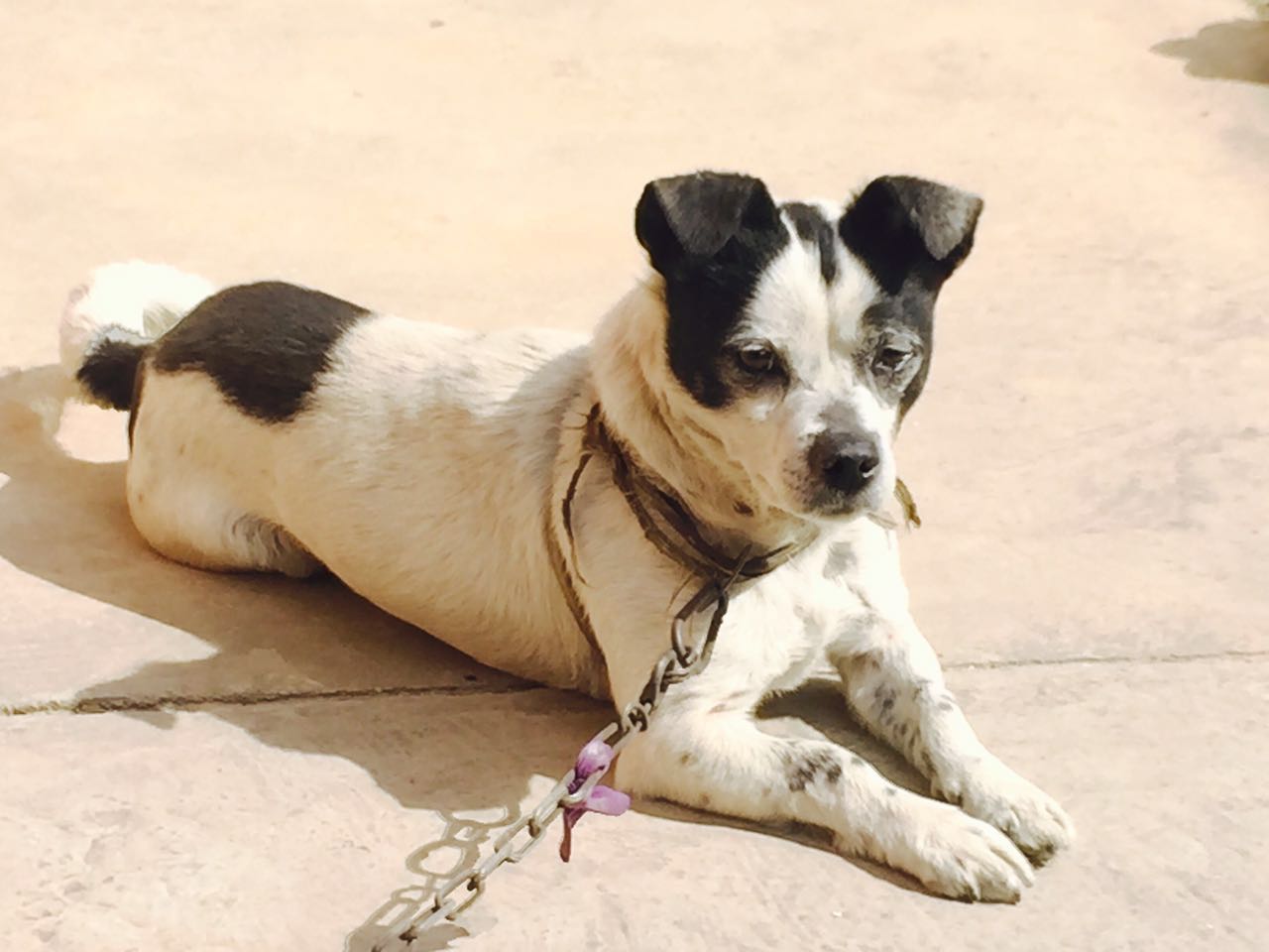
 | 品种:混种犬 |
| 年龄:未知 | |
| 性别:雄 | |
| 诊断:肱三头肌变异 | |
01 主诉及病史
在对其双侧前肢进行尸体解剖时,发现双侧肱三头肌都有一个额外的起源点。
该犬是从一家私人动物收容所获得的,用于教学目的。
02 检查
该犬外观正常,但体型较小,腿较短。根据美国兽医协会《动物安乐死指南》(2020 年版)的建议,通过静脉注射戊巴比妥钠(85 mg/kg)对该犬实施了安乐死。为了更好地观察动脉,在颈总动脉中注入了红色明胶。
03 解剖所见
肱三头肌的一个变异的附属头通过肌肉纤维从肱骨颈部的内侧起源,正好位于小结节的尾部。它的起始点位于肱骨冠状肌与肩胛下肌背侧终止点之间,然后向远端穿过,在插入处与内侧头的近半部尾侧融合(下图a-c)。因此,可以简单地将其视为肱三头肌的第二个附属头。左侧肢体的变异头要比右侧肢体的大(下图d)。左侧肢体的内侧头分为两个内侧和外侧腹部,而右侧则没有。左侧桡侧动脉从内侧头和变异头之间的远侧穿过,而右侧则从变异头的尾部穿过。沿其走向,该动脉向包括变异头在内的肱三头肌的肌肉头发出了数条分支。还发现桡神经从侧面穿过变异头和长头。变异头的神经支配通过桡神经的相同肌肉分支到达内侧头(下图b-d)。
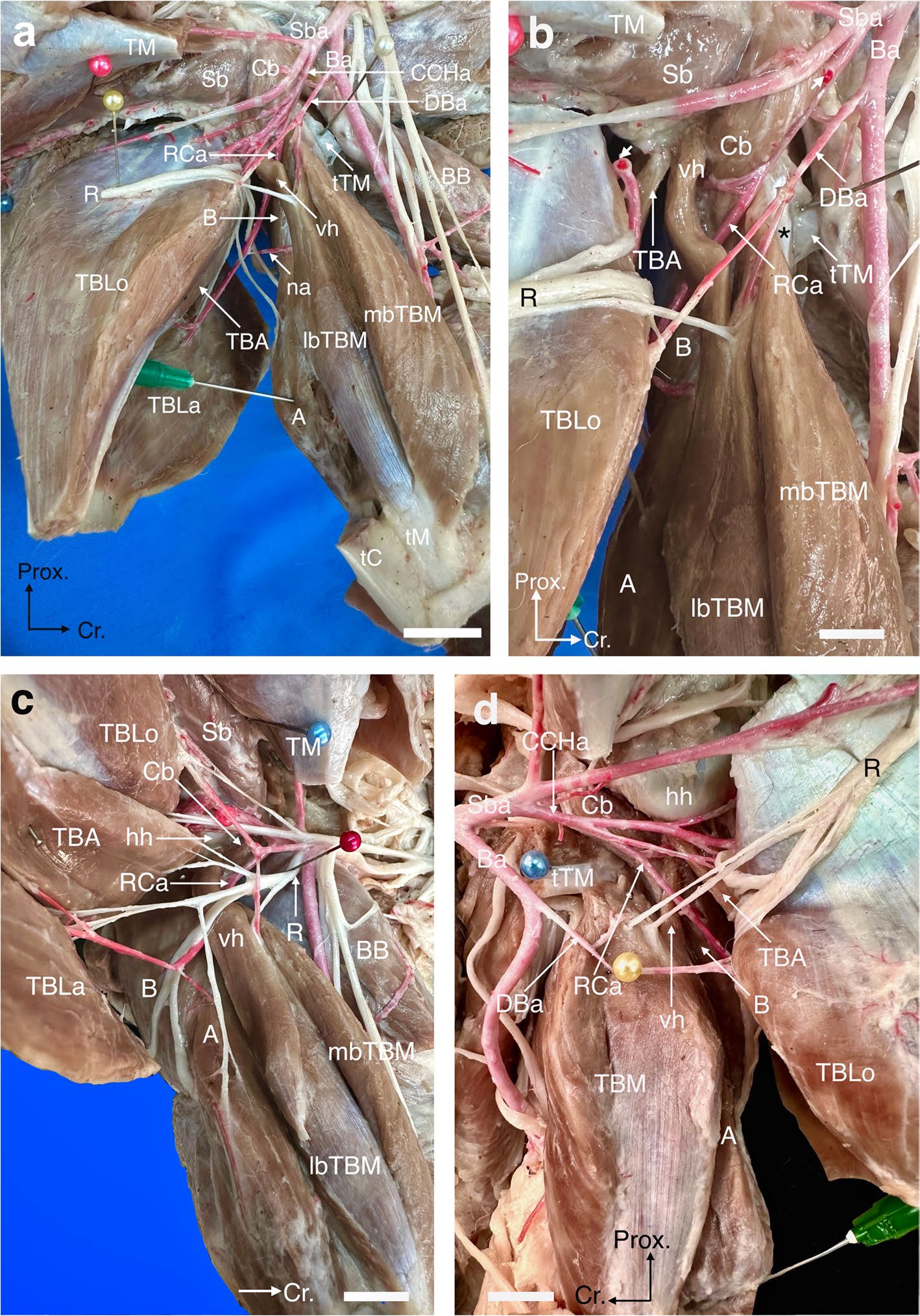
↑ (a)左肱内侧视图,显示肱三头肌变异头(vh)及相关结构。A,肘肌;B,肱肌;Ba,肱动脉;BB,肱二头肌;Cb,喙肱肌;CCHa,旋臂后动脉;RCa,桡动脉;DBa,肱深动脉;lbTBM,肱三头肌(内侧头)外侧腹肌群;mbTBM,肱三头肌(内侧头)内侧腹肌群;na,营养动脉;R,桡神经;Sb,肩胛下肌群;Sba,锁骨下动脉;tC,肱三头肌附属头(TBA)、外侧头(TBLa)和长头(TBLo)的共同肌腱插入部;tM:肱三头肌内侧头的肌腱插入部;TM:大圆肌及其肌腱插入部(tTM)。(b)左肱骨尾内侧,显示Sba肌肉分支(箭头所指)的附着位置。注意从肱三头肌变异头和内侧头之间穿过的RCa。*,TBM的肌腱起源。(c)左侧肱骨的尾内侧,显示vh与内侧头近半部尾侧的连接部位。hh,肱骨头。(d)右肱骨内侧视图,显示RCa与vh的位置关系。
在左侧肱骨中部发现的另一个变异是正中神经(M)与肌皮神经(MC)之间的细小沟通,该沟通贯穿肱动脉尾部(下图)。这种沟通与正中神经到肌皮神经的常规沟通方向相反。
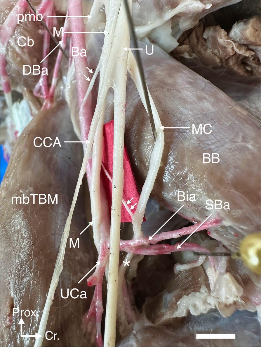
↑ 左肱内侧视图,显示正中神经(M)和肌皮神经(MC)之间的双向沟通(箭头和星号)。Ba,肱动脉;BB,肱二头肌;Bia,肱二头肌动脉;Cb,喙肱肌;CCA,尾侧皮前肱神经;UCa,尺侧副动脉;UCa,尺侧动脉;DBa,肱深动脉;mbTBM,肱三头肌(内侧头)内侧腹肌群;pmb,MC至BB的近端肌肉分支; SBa,肱浅动脉;U,尺神经。
04 讨论
家犬肱三头肌的常见解剖结构包括四个肌肉头:内侧头有一条明显的肌腱,来自肱骨小结节嵴上的一点,分别位于大圆肌和肱骨冠状肌插入部位的头部和尾部之间。它形成了一个狭窄的纺锤形肌腹,肌腹的末端是一条位于肩胛骨内侧的扁平肌腱。肱三头肌其他具有混合插入肌腱的头包括长头、外侧头和附属头[1-3]。
根据对野生食肉动物前肢肌肉组织的研究,某些物种的肱三头肌最多可由五个头组成,分别是:长头、大头、外侧头、角状头、附属头、内侧头和内侧附属头。最后一个位于内侧岬的远端[4-17]。既往研究在河狐中描述了四个头[5],而大多数犬科动物(如食蟹狐[11-12])都描述了相同数量的肌肉头。
最近一篇关于犬亚目的文章称,该亚目的大多数物种只有四个肌肉头,因为大多数作者命名的角状头和内侧附属头实际上分别是前肱筋膜张肌和内侧肌[10]。即使是Spoor和Badoux等人的研究也只描述了鬣羚的四个肌肉头[9],与家犬的四个肌肉头相似。
肱三头肌四个头的主要血液供应来自肱骨尾侧环动脉。其远端分支、桡侧动脉和伴行的桡神经在肱三头肌的大臂和背阔肌末端以及该肌肉的内侧头和附属头之间的远端运行[3]。
根据人类的报告,肱三头肌与其他肌肉相比很少出现变异[18],而据笔者所知,家犬尚未出现过这种肌肉变异的报告。本报告介绍了在一具成年雄性杂交家犬尸体上发现的肱三头肌双侧发育的额外肌肉头。
对其他食肉动物肱三头肌的研究表明,它可以由4-6个不同的头组成[4-17]。根据其他食肉动物肌肉图谱中肱骨三头肌的起源点,大多数食肉动物的肱骨三头肌起源于肱骨柄尾部表面的近端,包括肱骨颈[5, 6, 8, 10, 14, 15, 17, 19],本文中的变异头可能来自肱骨颈的内侧。
此外,桡侧动脉和肱三头肌之间的位置关系表明,在猎犬[3, 20]、食蟹狐[11]、蜜熊[10]、食蟹浣熊[19]和猫科动物[20]中,桡侧动脉穿过肱三头肌的外侧,到达肱三头肌的内侧。该动脉在这些食肉动物中的解剖关系也支持了该犬的额外头来自肱骨颈的假说。
总之,本文介绍的肱三头肌第二附属头的发展有助于理解食肉动物前肢肌肉进化过程中的系统发育和本体起源问题。
文献来源:Kamali Y. Five-headed triceps brachii muscle, with an unusual communication between the musculocutaneous and median nerves in a cross-breed dog cadaver: a case report. BMC Vet Res. 2025 Mar 3;21(1):130.
参考文献
1.Nickel R, Schummer A, Seiferle E, Frewein J, Wilkens H, Wille KH, Siller W, Stokoe W. The anatomy of the domestic animals. Berlin: Verlag Paul Parey. In: The locomotor system of the domestic mammal; 1986;1.
2.König HE, Hans-Georg HG, Bragulla H. Veterinary anatomy of domestic mammals: textbook and colour atlas. Berlin: Schattauer Verlag; 2007.
3.Hermanson JW, de Lahunta A. Miller and Evans’ anatomy of the dog – E-book. Berlin: Elsevier Health Sciences; 2018.
4.Ercoli MD, Álvarez A, Stefanini MI, Busker F, Morales MM. Muscular anatomy of the forelimbs of the lesser grison (Galictis cuja), and a functional and phylogenetic overview of Mustelidae and other Caniformia. J Mamm Evol. 2015;22(1):57–91.
5.de Souza JP, Santos LMRPD, Viotto-Souza W, de Carvalho NDC, Souza EC, Kasper CB, Abidu-Figueiredo M, Santos ALQ. Functional myology of the thoracic limb in Pampas fox (Lycalopex gymnocercus): a descriptive and comparative analysis. J Anat. 2018;233(6):783–806.
6.Dunn RH, Beresheim A, Gubatina A, Bitterman K, Butaric L, Bejes K, Kennedy S, Markham S, Miller D, Mrvoljak M, et al. Muscular anatomy of the forelimb of tiger (Panthera tigris). J Anat. 2022;241(1):119–44.
7.Fisher RE, Adrian B, Barton M, Holmgren J, Tang SY. The phylogeny of the red panda (Ailurus fulgens): evidence from the forelimb. J Anat. 2009;215(6):611–35.
8.Julik E, Zack S, Adrian B, Maredia S, Parsa A, Poole M, Starbuck A, Fisher RE. Functional anatomy of the forelimb muscles of the ocelot (Leopardus pardalis). J Mamm Evol. 2012;19(4):277–304.
9.Spoor C, Badoux D. Descriptive and functional morphology of the neck and forelimb of the striped hyena (Hyaena hyaena, L. 1758). Anat Anz. 1986;161(5):375–87.
10.Vélez-García JF, Blanco DAC, Gómez GM, Cañas SSM. Descriptive study of the intrinsic muscles of the shoulder and brachium in kinkajou (Potos flavus) and an evolutionary analysis within the suborder Caniformia. Vertebr Zool. 2023;73:957–80.
11.Vélez J, Ramírez J, Aristizábal O. An anatomic description of intrinsic brachial muscles in the crab-eating fox (Cerdocyon thous, Linnaeus 1776) and report of a variant arterial distribution. Anat Histol Embryol. 2018;47(2):180–3.
12.Pereira SG, Santos ALQ, Borges DCS, Queiroz PRR, Silva JORD. Anatomia óssea e muscular da escapula e braço de Chrysocyon brachyurus (Carnívora, Canidae). Cienc Anim Bras. 2016;17:622–32.
13.Barone R. Anatomie comparée des mammifères domestiques: Arthrologie et myologie. Berlin: Tome deuxième. ACV; 2020.
14.Smith HF, Townsend KEB, Adrian B, Levy S, Marsh S, Hassur R, Manfredi K, Echols MS. Functional adaptations in the forelimb of the Snow Leopard (Panthera uncia). Integr Comp Biol. 2021;61(5):1852–66.
15.Viranta S, Lommi H, Holmala K, Laakkonen J. Musculoskeletal anatomy of the Eurasian lynx, Lynx lynx (Carnivora: Felidae) forelimb: adaptations to capture large prey? J Morphol. 2016;277(6):753–65.
16.Smith HF, Adrian B, Koshy R, Alwiel R, Grossman A. Adaptations to cursoriality and digit reduction in the forelimb of the African wild dog (Lycaon pictus). PeerJ. 2020;8:e9866.
17.Tarquini J, Mosto MC, Ercoli MD. Functional and phylogenetic interpretation of the forelimb myology of two South American carnivorans, the ring-tailed coati (Nasua nasua) and crab-eating raccoon (Procyon cancrivorus). J Morphol. 2023;284(6):e21587.
18.Fabrizio PA, Clemente FR. Variation in the triceps brachii muscle: a fourth muscular head. Clin Anat (New York, NY). 1997;10(4):259–63.
19.Vélez García JF, Carrión Blanco DA, Moreno Gómez G, de Carvalho Barros RA, Miglino MA. Evolutionary derivation inferences of the intrinsic shoulder and brachial muscles in crab-eating raccoon (Procyon cancrivorus, Caniformia, Carnivora) based on the topology, innervation, and anatomical variants. Zoomorphology. 2024;143(3):795–818.
20.Barone R. Anatomie comparée des mammifères domestiques. Tome 5. In: Angiologie. Paris: Vigot; 1996.