| 一般情况 | |
|---|---|
 | 品种:混种犬 |
| 年龄:8岁 | |
| 性别:雄 | |
| 是否绝育:是 | |
| 诊断:右侧髌骨松弛 | |
01 主诉及病史
右后肢跛行。
自领养时(4年前)就出现后肢跛行。6个月前曾接受过评估,被诊断为右侧髌骨松弛。
02 检查
骨科检查发现该犬右后肢跛行2级,右侧髌骨松弛3级。体重45.6千克,肥胖,身体状况评分为9/9。
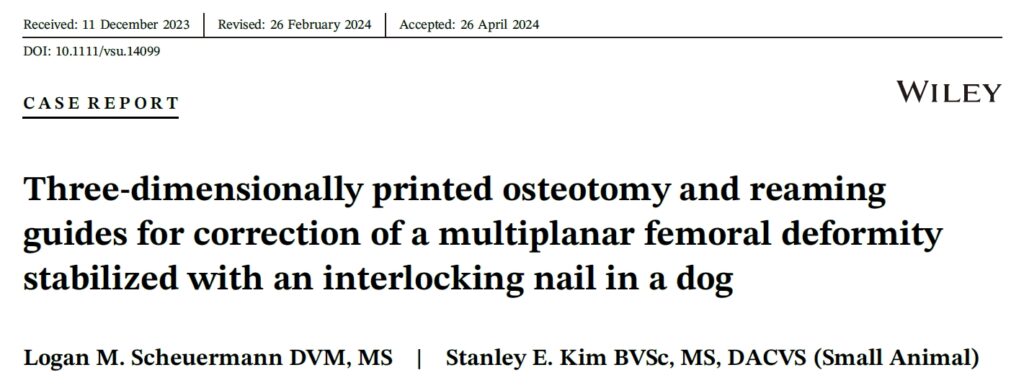
双侧股骨正侧位X光片显示双侧股骨外翻畸形,并伴有内翻和内扭转,疑似股骨连接不良(下图)。站立和行走时,右侧膝关节过度伸展。
为了制定手术计划,对双侧后肢进行了CT扫描,切片厚度为0.5 mm,切片重叠度为0.3 mm。将CT数据导入生物建模软件,通过测量每个股骨节段骨干线之间的轴向角度,确定每个节段的扭转对齐情况。结果发现,在旋转角中心近端,有14.8°的屈曲、10.3°的后凸和15.0°的外扭。在旋转角中心远端,有20.5°的内翻,62.9°的外翻和50.0°的内扭。
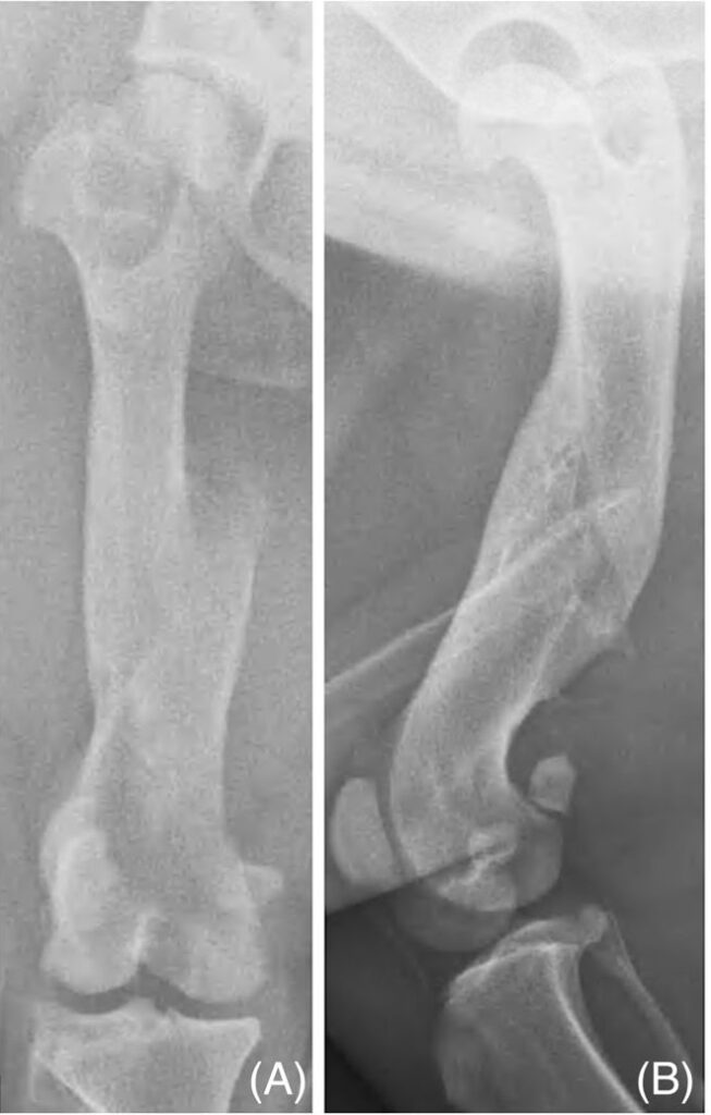
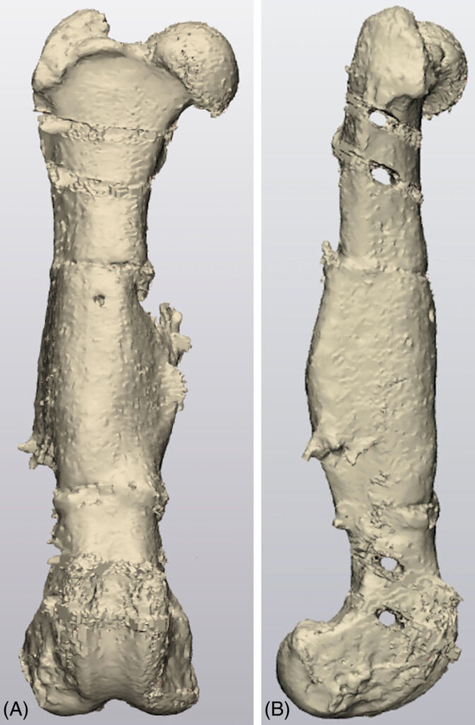
采用与后肢相同的技术对135 mm长、8 mm直径的交锁钉进行CT扫描,以创建一个虚拟的交锁钉,并将其导入到规划软件中。在软件中设计了定制的近端、中间和远端扩孔导向器,包括外侧钉套、符合股骨外侧形状的定制底座以及与股骨截骨表面相匹配的插管扩孔套(下图)。基座设计有0.3 mm的间隙,以考虑到骨膜和区域软组织。
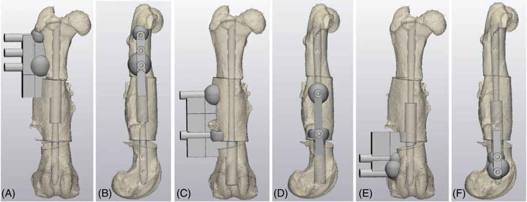
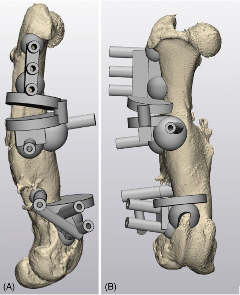
定制的近端和远端截骨导板的设计符合每个股骨形态(下图)。两个截骨导板都由可容纳3.2 mm半针的针套、截骨架和定制的底座组成,底座与针套和截骨架之间的间隙为0.3 mm。最后,使用立体光刻3D打印机用生物相容性树脂进行3D打印。
03 手术
3D打印的骨模型和定制手术导板处理和固化后进行蒸汽灭菌。手术在初次评估72天后进行。
麻醉后使用右美托咪定(0.22 μg/kg)和布比卡因(0.33 mg/kg)对右髂前股神经和坐骨旁神经进行阻滞。麻醉诱导后注射头孢唑啉(22 mg/kg IV),术中每隔90分钟注射一次。在狗处于背外侧斜卧位的情况下,进行了右股骨外侧入路和右膝关节外侧髌旁入路。
将截骨导板安装到最合适的位置,并用”1/8″Steinmann钉固定(下图A)。进行近端和远端截骨后,取下截骨导板,但不移除Steinmann钉。将近端、中部和远端扩孔导板套在Steinmann钉上,用4.5 mm钻头逆行扩孔髓管(下图B)。
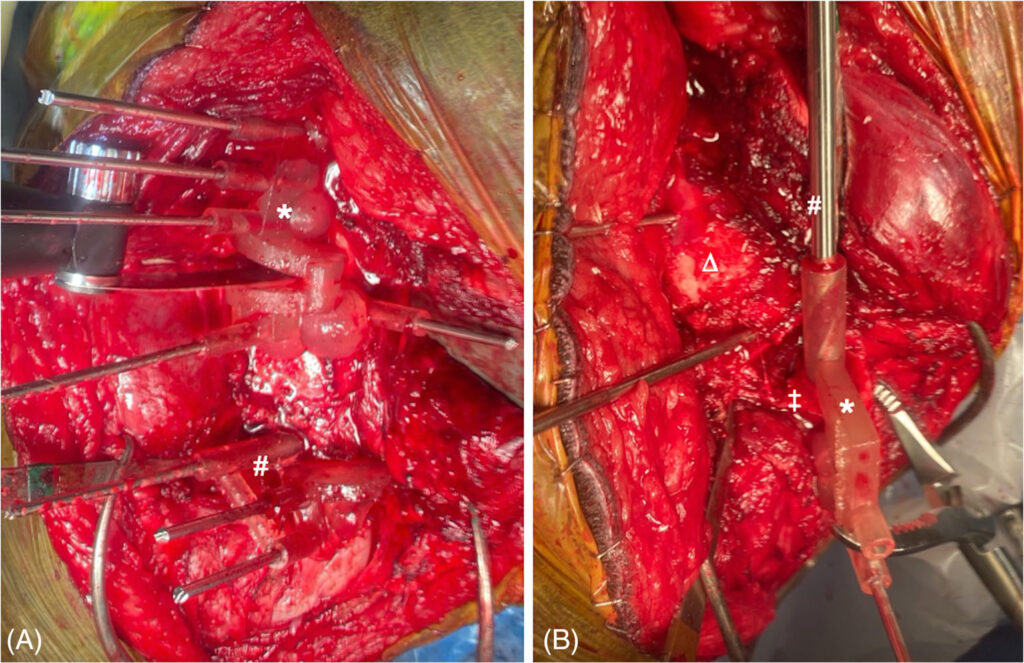
在扩孔的同时,依次拔出Steinmann钉,以方便对髓腔进行扩孔。取出扩孔导板。然后用锥子进一步制备每个节段的髓腔,并将交锁钉以顺行方式插入近端和中间节段,然后打入股骨远端节段。术中通过透视评估截骨缩小情况。
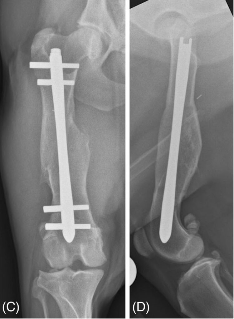
将关节镜针插入Steinmann钉创建的骨道的股骨皮质,分别使用4.3 mm和3.2 mm钻头对钉道的顺侧和横侧皮质进行钻孔,然后插入四个锁定螺栓。在远端开口楔形截骨术中插入了近端截骨术中切除的楔形皮质。术后拍摄了右股骨X光片(下图),手术总时间为149分钟。
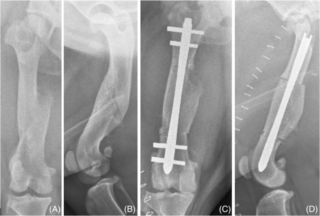
术后,该犬接受了头孢唑啉(22 mg/kg IV)治疗,每8小时一次,持续24小时,之后转为头孢氨苄(22 mg/kg PO),每12小时一次,卡泊芬净(2.2 mg/kg PO),芬太尼恒速输注(2-3 μg/kg/h IV),持续18小时,加巴喷丁(13 mg/kg PO),每8小时一次。
04 预后
术后1天,该犬右后肢无负重跛行,右侧大腿软组织轻度肿胀。在整个膝关节活动范围内,右侧髌骨都在膝关节沟内。
术后1周和3周,该犬右后肢间歇性无负重跛行。在整个膝关节活动范围内,髌骨都位于膝关节沟内。
术后股骨长度比虚拟平面图短0.7 mm(下图),比术前股骨长1.2 mm。术后股骨长度比对侧股骨短20.4%。
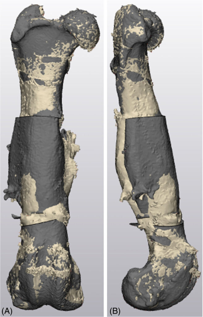
校正后的股骨与术前相比,股骨曲度增加了17.7°。与对侧股骨和模拟结果相比,术后曲度分别增加了3.8°和2.2°。术后矢状面对位与虚拟计划对位在0.4°以内,复位增加。矫正后的股骨比术前股骨的后凸减少了17.5°,比对侧股骨的后凸增加了11.4°。术后股骨的扭转分别比虚拟计划的股骨和对侧股骨大1.6°和2.8°(即更多前翻),比术前股骨小17.2°(即更多正常翻转)。
术后2个月,该犬的目视步态检查结果良好,髌骨在整个膝关节活动范围内都能在膝关节沟内,无移位。术后两个月的正位X光片和CT 扫描(下图)显示股骨对齐、植入物良好。
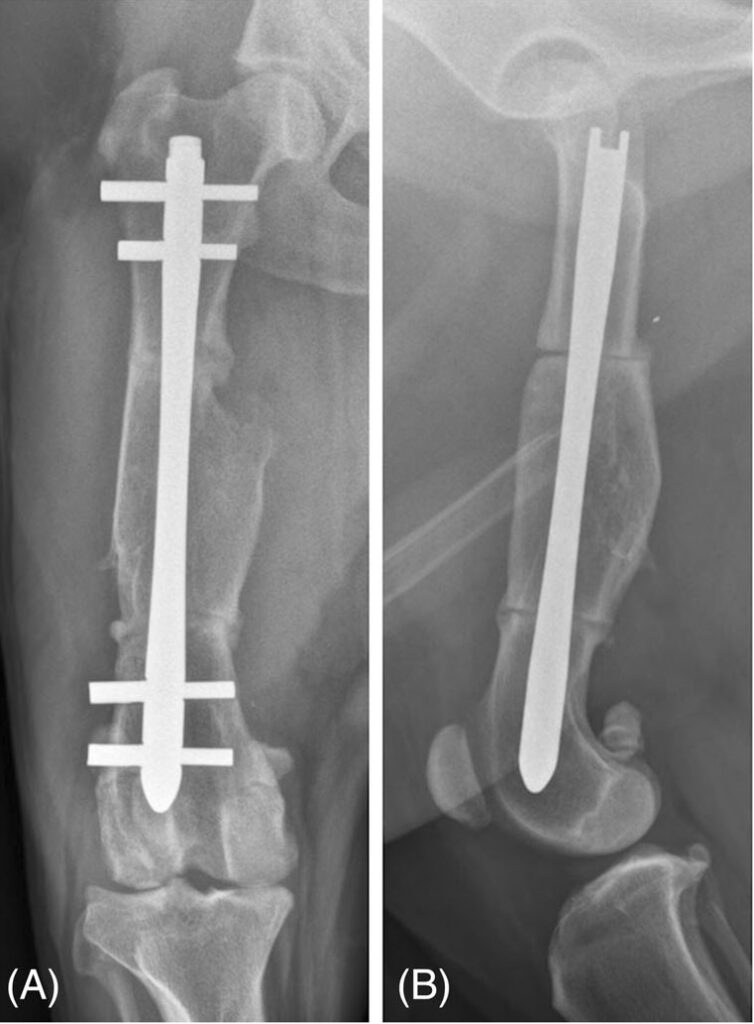
术后10个月评估,该犬的跗骨过度伸展,与术前检查结果相当。髌骨在膝关节沟内的运动正常,没有脱位。右侧股骨正侧位X光片显示,截骨处皮质连续,没有明显的截骨线(下图)。交锁钉和螺栓稳定,跗骨正侧位X光片无异常。
05 讨论
髌骨脱位与后肢的先天、发育或外伤异常有关,会导致股四头肌功能受损,并可能造成跛行[1,2]。与滑车成形术和胫骨结节移位术相比,较高程度的髌骨脱位常伴有股骨畸形,更有可能需要进行矫正性截骨术,以降低术后再脱位的风险[1,3,4]。
使用X光片对股骨畸形进行评估具有挑战性,尤其是对于伴有扭转异常的多平面畸形[4-6]。因此,CT通常是首选的成像方式,因为它比X光片能更准确地量化三维结构[7,8]。
股骨远端截骨术是治疗髌骨脱位和并发股骨畸形的有效方法,尤其适用于正面畸形[3,4]。最近,虚拟手术规划和3D打印定制手术导板被用于狗的股骨远端截骨术[9]。在一项研究中,3D打印股骨远端截骨和还原导板有助于在术前计划的2.3°和1.7°范围内分别矫正股骨远端内翻和股骨扭转[9]。
使用交锁钉矫正畸形可能具有优势,因为交锁钉在生物力学上非常坚固,理论上有利于轴向对齐[10-13]。以前曾有人描述过用交锁钉矫正股骨远端畸形的方法[10,14,15],但该手术在技术上可能比较困难,尤其是对复杂的畸形。
在置入交锁钉的过程中,准确置入螺栓是一项挑战,尤其是远端螺栓[10,16]。虽然3D打印导板可以生成精确的股骨远端截骨,并用骨板进行稳定,但传统的导板设计可能不适合与交锁钉一起使用。
本病例报告描述了利用虚拟手术规划和3D打印定制的截骨、钻孔和扩孔导板,成功矫正了一只狗的双侧多平面股骨畸形。术后股骨长度以及正面、矢状面和轴面的对位与虚拟规划的股骨模型分别相差0.7 mm、2.2°、0.5° 和 1.6°。如果采用传统的术前规划和术中技术,要达到这样的精确度可能更具挑战性。
该病例的结果与之前关于使用定制3D打印导板对狗进行畸形矫正的报告结果一致。一项前瞻性研究报告了利用定制的截骨和还原导板对10只患有髌骨内侧松弛的狗进行了准确的股骨远端截骨术,并用锁定钢板进行了稳定[9]。另一项研究也报告了利用定制手术导板对肱骨前端畸形进行精确矫正的结果,桡骨正面、矢状面和轴向的平均角度与实际计划的对齐角度分别在1.8°、2.5°和3.5°以内[27]。
使用交锁钉稳定股骨畸形矫正的方法此前已有描述[10,14,15]。本病例选择交锁钉有多种原因。将交锁钉放置在髓管内有利于正面的重新调整。股骨截骨术通常使用钢板进行稳定,植入失败的情况并不常见[3,4,30],但从生物力学角度来看,交锁钉比偏心放置的钢板更有利,因为交锁钉更靠近股骨中轴,可将负重时的弯曲力矩降至最低。
股骨近端和远端螺栓位于股骨干骺端,因此只需一个植入物即可弥合多处截骨,而钢板固定则可能需要多个植入物。股骨远端钢板必须放置在膝关节囊附近或上方,这可能会导致局部刺激[3,30]。
总之,利用虚拟手术规划和3D打印定制截骨和扩孔导板有助于准确矫正股骨畸形和放置交锁钉。体内外研究和临床研究可能有助于进一步评估定制扩孔导板在矫正肢体成角畸形过程中放置交锁钉的准确性。
文献来源:Scheuermann LM, Kim SE. Three-dimensionally printed osteotomy and reaming guides for correction of a multiplanar femoral deformity stabilized with an interlocking nail in a dog. Vet Surg. 2024 May 6.
参考文献
1. Perry KL, Déjardin LM. Canine medial patellar luxation. J Small Anim Pract. 2021;62(5):315-335.
2. Roy RG, Wallace LJ, Johnston GR, Wickstrom SL. A retrospective evaluation of stifle osteoarthritis in dogs with bilateral medial patellar luxation and unilateral surgical repair. Vet Surg. 1992;21(6):475-479.
3. Brower BE, Kowaleski MP, Peruski AM, et al. Distal femoral lateral closing wedge osteotomy as a component of comprehensive treatment of medial patellar luxation and distal femoral varus in dogs. Vet Comp Orthop Traumatol. 2017;30(1):20-27.
4. Swiderski JK, Palmer RH. Long-term outcome of distal femoral osteotomy for treatment of combined distal femoral varus and medial patellar luxation: 12 cases (1999–2004). J Am Vet Med Assoc. 2007;231(7):1-6.
5. Jackson GM, Wendelburg KL. Evaluation of the effect of distal femoral elevation on radiographic measurement of the anatomic lateral distal femoral angle. Vet Surg. 2012;41(8):994-1001.
6. Adams RW, Gilleland B, Monibi F, Franklin SP. The effect of valgus and varus femoral osteotomies on measures of anteversion in the dog. Vet Comp Orthop Traumatol. 2017;30(3):184-190.
7. Dudley RM, Kowaleski MP, Drost WT, Dyce J. Radiographic and computed tomographic determination of femoral varus and torsion in the dog. Vet Radiol Ultrasound. 2006;47(6):546-552.
8. Yasukawa S, Edamura K, Tanegashima K, et al. Evaluation of bone deformities of the femur, tibia, and patella in toy poodles with medial patellar luxation using computed tomography. Vet Comp Orthop Traumatol. 2016;29(1):29-38.
9. Hall EL, Baines S, Bilmont A, Oxley B. Accuracy of patientspecific three-dimensional-printed osteotomy and reduction guides for distal femoral osteotomy in dogs with medial patella luxation. Vet Surg. 2019;48(4):584-591.
10. Déjardin LM, Perry KL, von Pfeil DJF, Guiot LP. Interlocking nails and minimally invasive osteosynthesis. Vet Clin North Am Small Anim Pract. 2020;50(1):67-100.
11. Déjardin LM, Lansdowne JL, Sinnott MT, Sidebotham CG, Haut RC. In vitro mechanical evaluation of torsional loading in simulated canine tibiae for a novel hourglass-shaped interlocking nail with a self-tapping tapered locking design. Am J Vet Res. 2006;67(4):678-685.
12. Lansdowne JL, Sinnott MT, Déjardin LM, Ting D, Haut RC. In vitro mechanical comparison of screwed, bolted, and novel interlocking nail systems to buttress plate fixation in torsion and mediolateral bending. Vet Surg. 2007;36(4):368-377.
13. Déjardin LM, Guillou RP, Ting D, Sinnott MT, Meyer E, Haut RC. Effect of bending direction on the mechanical behaviour of interlocking nail systems. Vet Comp Orthop Traumatol. 2009;22(4):264-269.
14. Witte PG, Scott HW. Treatment of lateral patellar luxation in a dog by femoral opening wedge osteotomy using an interlocking nail. Vet Rec. 2011;168(9):243.
15. Guillou RP, Guiot LP, Déjardin LM. Angle-stable interlocking nail fixation for distal femoral corrective osteotomy associated with medial patellar luxation. Vet Comp Orthop Traumatol. 2013;26(4):A11.
16. Duhautois B. Use of veterinary interlocking nails for diaphyseal fractures in dogs and cats: 121 cases. Vet Surg. 2003;32(1):8-20.
17. Quinn MM, Keuler NS, Lu Y, Faria MLE, Muir P, Markel MD. Evaluation of agreement between numerical rating scales, visual analogue scoring scales, and force plate gait analysis in dogs. Vet Surg. 2007;36(4):360-367.
18. Paley D, Herzenberg JE, Tetsworth K, McKie J, Bhave A. Deformity planning for frontal and sagittal plane corrective osteotomies. Orthop Clin North Am. 1994;25(3):425-465.
19. Wei F, Li J, Zhou C, et al. Combined application of singleenergy metal artifact reduction and reconstruction techniques in patients with cochlear implants. J Otolaryngol Head Neck Surg. 2020;49(1):1-9.
20. Yasaka K, Kamiya K, Irie R, Maeda E, Sato J, Ohtomo K. Metal artefact reduction for patients with metallic dental fillings in helical neck computed tomography: comparison of adaptive iterative dose reduction 3D (AIDR 3D), forward-projected modelbased iterative reconstruction solution (FIRST) and AIDR 3D with single-energy metal artefact reduction (SEMAR). Dentomaxillofacial Radiol. 2016;45(7):1-7.
21. Kawahara D, Ozawa S, Yokomachi K, et al. Metal artifact reduction techniques for single energy CT and dual-energy CT with various metal materials. BJR Open. 2019;1(1):1-7.
22. Formlabs. Form Cure Manual. 2019. Accessed July 9, 2021.
23. Tomlinson J, Fox D, Cook JL, Keller GG. Measurement of femoral angles in four dog breeds. Vet Surg. 2007;36(6):593-598.
24. Peterson JL, Torres BT, Hutcheson KD, Fox DB. Radiographic determination of normal canine femoral alignment in the sagittal plane: a cadaveric pilot study. Vet Surg. 2020;49(6):1230-1238.
25. Serck BM, Karlin WM, Kowaleski MP. Comparison of canine femoral torsion measurements using the axial and biplanar methods on three-dimensional volumetric reconstructions of computed tomography images. Vet Surg. 2021;50(7):1518-1524.
26. Scheuermann LM, Kim SE, Lewis DD, Johnson MD, Biedrzycki AH. Minimally invasive plate osteosynthesis of femoral fractures with 3D-printed bone models and custom surgical guides: a cadaveric study in dogs. Vet Surg. 2023;52(6):827-835.
27. De Armond CC, Lewis DD, Kim SE, Biedrzycki AH. Accuracy of virtual surgical planning and custom three-dimensionally printed osteotomy and reduction guides for acute uni- and biapical correction of antebrachial deformities in dogs. J Am Vet Med Assoc. 2022;1-9:1-9.
28. Silveira F, Monotti IC, Cronin AM, et al. Outcome following surgical stabilization of distal diaphyseal and supracondylar femoral fractures in dogs. Can Vet J. 2020;61(10):1073-1079.
29. Coutin JV, Lewis DD, Kim SE, Reese DJ. Bifocal femoral deformity correction and lengthening using a circular fixator construct in a dog. J Am Anim Hosp Assoc. 2013;49(3):216-223.
30. Kim SE, Lewis DD. Corrective osteotomy for procurvatum deformity caused by distal femoral physeal fracture malunion stabilised with string-of-pearls locking plates: results in two dogs and a review of the literature. Aust Vet J. 2014;92(3):75-80.